Author: Alina Yang
Editors: Ethan Tai, Ian Cho
Artist: Emily Hu
In ancient times, people primarily believed that the inside of a human body consisted of a network of interconnected tubes and chambers. Due to religious and cultural boundaries, dissection was largely prohibited in many societies; cutting open a dead body was seen as a desecration. This forced early anatomists to extrapolate animal dissections to people, leading to inaccurate assumptions about human anatomy. One such primitive model was that of Hippocrates; he believed that the four humors — phlegm, black (melancholic), red (sanguine), and yellow (choleric) bile — determined a person’s temperament and an imbalance led to certain illnesses dependent on the excess or deficit of humors. In modern times, the change of societal norms and vast developments in technology enable new insights into the biological underpinnings of those ancient theories by allowing researchers to investigate bodily systems and functions safely.
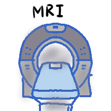
One such technology is Magnetic Resonance Imaging (MRI). As denoted by its name, at the heart of every MRI machine is a giant magnet. These magnets, weighing several tons and possessing exceptionally strong magnetic field strength, are measured in tesla — units of magnetic induction or magnetic flux density. Most clinical MRI machines range between 1.5 Tesla (T) and 3 Tesla, but some newer models have field strengths as high as 7 Tesla or more. In September 2021, the 11.7 Tesla MRI of the Iseult project was unveiled.
The magnet capitalizes on the properties of atomic nuclei. Like shaking a box of compasses until all the needles point in the same direction, when a patient lies down (or stands up) in the scanner, the magnet aligns the protons in their body. When a magnetic field is employed to align the typically randomly oriented protons within the tissue's water nuclei, an external Radio Frequency (RF) energy is introduced to the process. The nuclei then return to their resting alignment through relaxation processes and, in doing so, emit RF energy. The signals emitted from the initial RF are measured, and Fourier transformation is then used to convert the frequency information contained in each signal from the location in the scanned plane to corresponding intensity levels, which is displayed as the various shades of gray seen in MRI photographs.
Recently, a type of MRI known as diffusion tensor imaging (DTI) has emerged, making it possible to evaluate the organization of white matter, neuronal fibers in the brain that help the body process information. With this novel technique, the movement of water molecules in the brain and body can be detected with such precision that they can reveal the presence of a disease before symptoms can even appear by revealing tissue microstructural changes. Therefore, techniques such as DTI have become especially useful in studying neurological disorders like Alzheimer’s, multiple sclerosis, and even early signs of brain trauma, providing a glimpse into changes invisible to other imaging methods. This groundbreaking novelty is an example of how Magnetic Resonance Imaging (MRI) can extend beyond just capturing images of the body. With the emergence of a new technological era, MRIs and other radiologic modalities are evolving into diagnostic powerhouses capable of detecting subtle changes that make it possible to redefine early disease intervention. MRIs don’t just offer a snapshot of the present, but they also provide clues about the future trajectory of healthcare.
As illustrated by the DTI, the most fascinating aspect of MRIs is their customizability. By making small changes to the settings, you can focus on different tissues and conditions noted by the patient and/or previous medical history. Various types of images are created by varying the sequence of RF pulses applied and collected. Repetition time (TR), the amount of time between each successive pulse sequence, and time to echo (TE), the time between the delivery of the RF pulse and the recipient of the echo signal, can be varied to produce T1 and T2 images.

Tissues can be categorized by two relaxation times; T1 and T2. T1 is the longitudinal relaxation time, which is the time constant that determines the rate at which excited protons return to equilibrium. T2 is the transverse relaxation time, which is the time constant that determines the rate at which excited protons reach equilibrium or go out of phase with one another. T1-weighted images, produced by short TE and TR times, highlight fat and provide sophisticated anatomical detail, making it easy to distinguish between gray and white matter in the brain and throughout the body. T2-weighted images, produced by longer TE and TR times, primarily focus on fluid detection, ideal for detecting swelling, inflammation, or cysts.
Due to their relative novelty, MRIs aren’t perfect and have considerable room for improvement. MRIs are contraindicated due to their powerful magnetic interactions with metallic objects such as surgical metal implants, pacemakers, or cochlear implants. Since a single scan can take anywhere from 15 minutes to over an hour, depending on the type of scan ordered by the referring physician, patients with claustrophobia and a fear of tight spaces may find lying in a tight, noisy tube for up to an hour triggering. Regardless, MRIs' unparalleled detail and diagnostic precision make it an optimal choice for many patients.

MRI technology is continuously evolving, unlocking more possibilities for diagnostic radiology. Researchers are pushing the boundaries with ultra-high-field MRIs for better resolution and faster imaging techniques to reduce scan times for patients and increase technician efficiency. Beyond diagnostics, MRIs also aid scientists in studying everything from how the brain changes in Alzheimer’s to the performance of new cancer treatments at a microscopic level.
MRIs have become a keystone in modern healthcare, allowing doctors to see inside the human body with remarkable clarity that was once unimaginable, unraveling biological mysteries hidden in plain sight. As we continue to push the boundaries of what is possible with MRIs, the future holds incredible promise.
Citations: CEA. “The Most Powerful MRI Scanner in the World Delivers Its First Images!” CEA/English
Portal, 7 Oct. 2021, www.cea.fr/english/Pages/News/premieres-images-irm-iseult-
Kawahara, Daisuke, and Yasushi Nagata. “T1-Weighted and T2-Weighted MRI Image
Synthesis with Convolutional Generative Adversarial Networks.” Reports of Practical
Oncology and Radiotherapy, vol. 26, no. 1, 25 Feb. 2021, pp. 35–42,
National Institute of Biomedical Imaging and Bioengineering. “Magnetic Resonance
Imaging (MRI).” National Institute of Biomedical Imaging and Bioengineering, 17 July 2019,
NHS. “Overview - MRI Scan.” NHS, NHS, 26 July 2022, www.nhs.uk/conditions/mri-scan/.
Radiology (ACR), Radiological Society of North America (RSNA) and American
College of. “Magnetic Resonance Imaging (MRI) Procedures.” Radiologyinfo.org,
“Wellstar Health System.” Wellstar.org, 2025, www.wellstar.org/for-providers/request-
imaging-services/mri-of-the-liver. Accessed 26 Jan. 2025.
Comments